Every IVUS Frame in Its Place: Registering Intravascular Ultrasound to the Angiogram in Your Browser
Preprint status: This paper has been submitted to arXiv and is still in moderation. This post is a preview. We’ll add the arXiv link when it goes live.
Try it now: menonpg.github.io/ivus-xa-registration/?demo. One click loads a real, de-identified Left Main case.
The problem: the registration happens in the cardiologist’s head
Intravascular ultrasound (IVUS) shows what the coronary angiogram can’t: plaque composition, calcium arcs and stent apposition. The angiogram is the map, though. It’s the silhouette the interventionalist actually steers by.
Matching each IVUS cross-section to its spot on that map is still mostly done mentally. Commercial co-registration exists, but it’s tied to vendor consoles and has to be set up while the images are being acquired. Archived pullbacks, which hospitals have stored for years, can’t be co-registered after the fact. As a result, paired IVUS–angiogram data for training AI models is still scarce.
This paper, written with Dr. Srilakshmi Adhyapak (Cardiology, St. John’s Medical College Hospital, Bengaluru), describes a tool that registers archived cases in any modern browser.
What the tool does
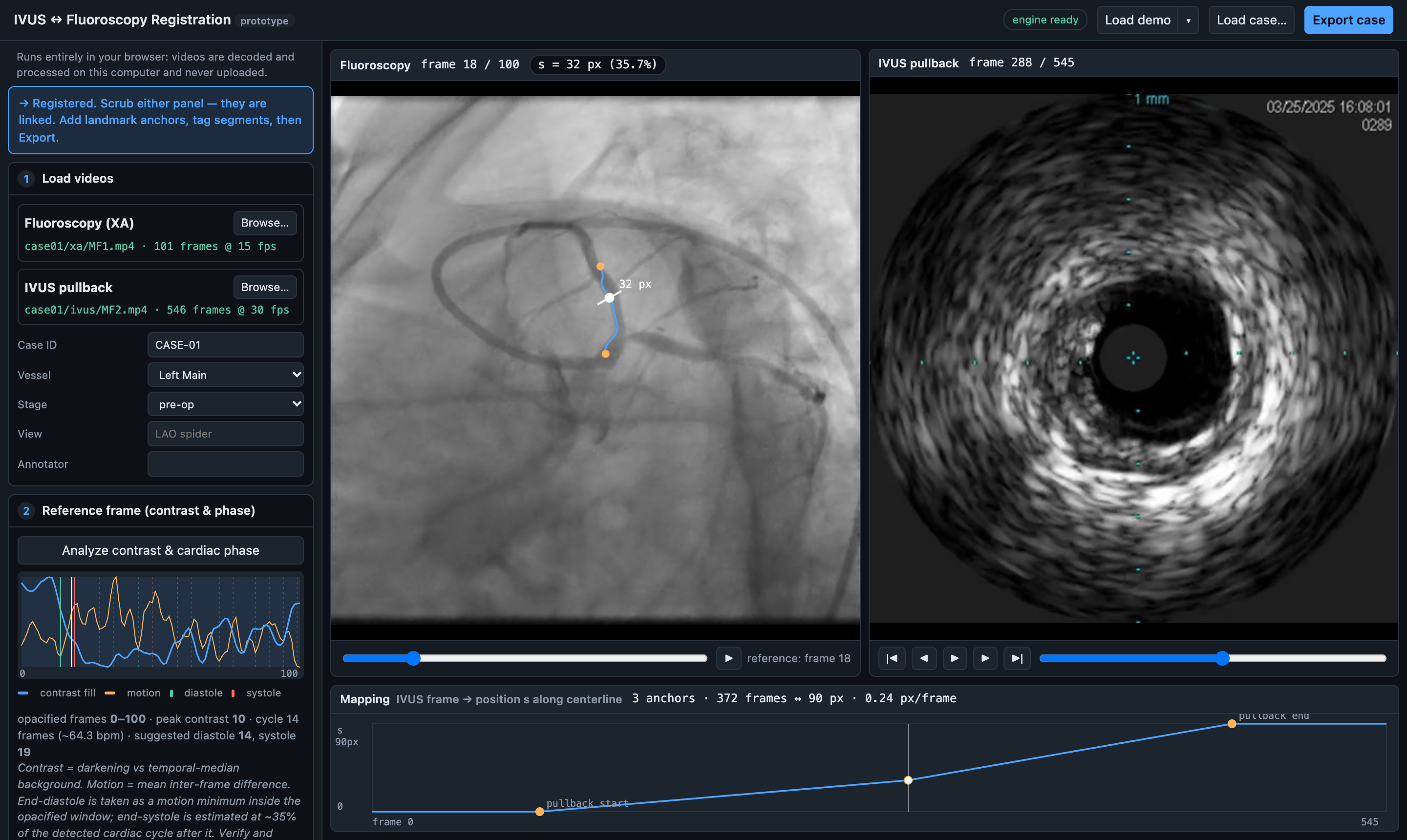
The workflow has five steps. The software proposes, and the clinician decides:
- Load videos. Open a folder with the angiographic run and the IVUS pullback. Each run gets a thumbnail and labels such as “pullback” or “short run”, so the right one is easy to find.
- Reference frame. The tool plots contrast filling and frame-to-frame motion, then suggests end-diastolic and end-systolic frames. Exported videos don’t carry an ECG, so this step relies on heuristics.
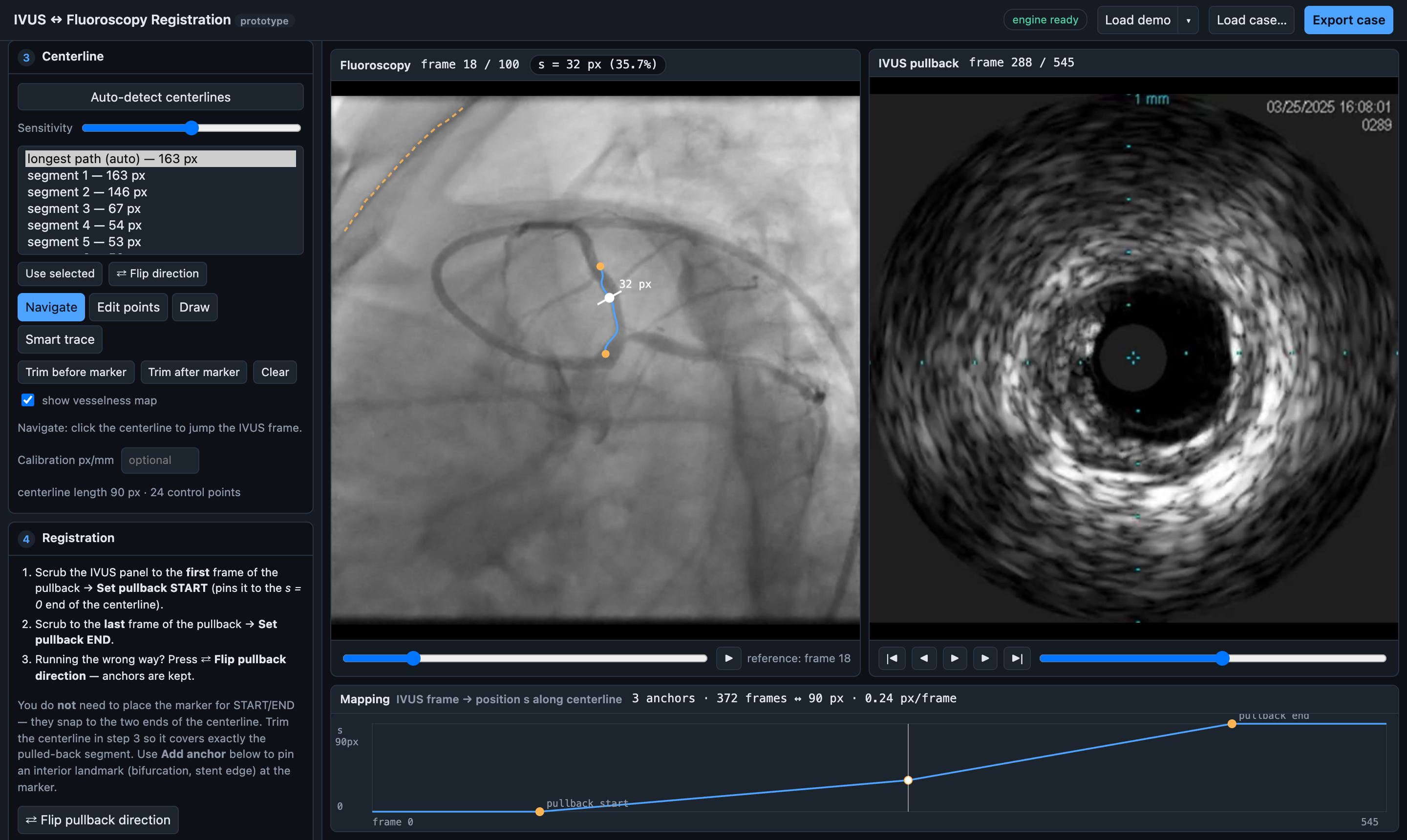
- Centreline. Frangi vesselness and skeleton analysis produce candidate centrelines. With smart trace, you click twice and a minimal-cost path follows the vessel between the two points.
- Registration. Pin the pullback’s START and END, then add landmarks such as side-branch ostia or stent edges. A monotone piecewise-linear map places every IVUS frame on the vessel.
- Tag and export. Tag frame ranges (calcium, lipid, stent, bifurcation…) and export a frame-level training record.
No install, no upload
All of this runs in the browser. The original Python analysis (numpy, scipy, scikit-image) runs unchanged in WebAssembly through Pyodide. Videos are decoded frame-accurately with WebCodecs. Patient videos stay on the local machine.
That has a practical consequence. To review a case, a clinician needs only a link and a folder of exported videos. There’s no IT ticket and no server to approve.
What we found
- The port is exact. On 6 angiographic runs, the in-browser analysis was bit-identical to the server pipeline when given identical frames. Browser decoding differed from ffmpeg by at most 2 grey levels. The suggested reference frames agreed in all 6 runs.
- It’s fast enough for interactive use. A 546-frame pullback decoded in 5.3 s. The one-click demo loads in about 6 s.
- The automatic steps are useful starting points, but the clinician still has to decide. On 36 phantoms with known ground truth, the clinician-guided smart trace stayed inside the lumen along 87–99% of the vessel. The fully automatic “longest path” centreline fell from 83% to 0% as noise rose and contrast dropped. On the real case, it followed the guiding catheter.
- Landmarks matter. In the registered Left Main case, adding one side-branch anchor moved mid-pullback positions by 17.5% of the segment compared with the usual constant-speed assumption. The probe doesn’t move at a constant speed along the vessel.
The bigger idea: virtual IVUS
Every registered case pairs each IVUS frame with an angiographic location, the local vessel image and the cardiac phase. The registration is the label, and that’s exactly the supervision cross-modal models need.
The paper lays out where AI can take over each step: learned phase detection, coronary segmentation in place of vesselness, automatic landmark proposals, and IVUS plaque segmentation. The most ambitious target is virtual IVUS: predicting the IVUS cross-section, or simpler targets like calcium presence, calcium arc or plaque burden, from the angiogram alone. That would be similar to how angiography-derived FFR replaced a pressure wire for many patients.
Nobody yet knows whether the angiogram contains enough information to do this. This tool makes the question testable, and the models can run in the browser through ONNX Runtime Web, so data still never has to leave the machine.
Try it
- Live tool + demo: menonpg.github.io/ivus-xa-registration/?demo
- Paper: Every IVUS Frame in Its Place: A Zero-Install Browser Tool for Registering Intravascular Ultrasound to the Coronary Angiogram, and a Path to Angiography-Only Plaque Imaging. Prahlad G. Menon, Srilakshmi Adhyapak. arXiv link coming soon.
This is a research tool and not a medical device.